##
“Neurocutaneous markers” are visible skin findings that signal an underlying **neurocutaneous (phakomatosis) syndrome** with a known risk of brain-spinal cord malformations (for example, tuberous sclerosis, NF1).
In contrast, **ASD (autism spectrum disorder)** is a neurodevelopmental diagnosis defined by social-communication differences and restricted-repetitive behaviors, **not** by a specific skin lesion.
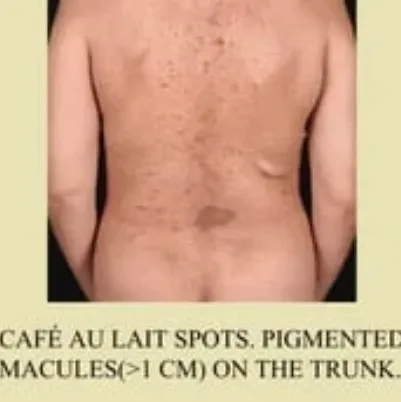
Research does show that some **congenital-nevoid skin markers** (especially café‑au‑lait macules and hyperpigmented pigmentary mosaicism) occur more often in children with ASD than in controls, but **they are not specific or strong enough to diagnose ASD**; they simply raise clinical suspicion and prompt closer developmental monitoring.
***
## What “neurocutaneous markers” usually means
In paediatrics and neurology, “neurocutaneous markers” typically refers to **cutaneous stigmata that predict structural CNS-spinal anomalies or neurocutaneous syndromes**, such as:
- Midline lumbosacral dimples, hairy patches, or vascular anomalies → risk of **occult spinal dysraphism** (tethered cord, lipomyelomeningocele, etc.).
- Characteristic lesions of **neurocutaneous syndromes**:
- **Tuberous sclerosis complex (TSC)**: ash‑leaf hypopigmented macules, facial angiofibromas, shagreen patch.
- **Neurofibromatosis type 1 (NF1)**: multiple café‑au‑lait macules, axillary-inguinal freckling, neurofibromas.
- **Sturge–Weber syndrome**: port‑wine stain (facial capillary malformation) in a V1 distribution.
These markers matter because they often **trigger imaging, specialist referral, and surveillance** for seizures, developmental delay, or other neurological issues.
***
## ASD: a neurodevelopmental, not neurocutaneous, diagnosis
ASD is diagnosed **behaviorally**, using developmental history and observation (e.g., DSM‑5‑TR-ICD‑11 criteria), not by a pathognomonic lab test or skin sign.
Key points:
- No single **laboratory, genetic, imaging, or dermatological test** is currently approved to diagnose ASD.
- Biomarker research (EEG, eye‑tracking, MRI, blood-urine markers) is active but **not yet ready for routine clinical diagnosis**.
***
## Where the two overlap: skin markers studied in ASD
Several studies have asked: *Do children with ASD have more congenital-nevoid skin lesions than other children?* Findings (mostly from Indian cohorts) suggest:
- **Higher prevalence of congenital-nevoid lesions in ASD** vs controls (odds ratio ≈ 3.12).
- Most associated lesions:
- **Hyperpigmented pigmentary mosaicism** (Blaschko‑linear-segmental patterns): OR ≈ 2.76.
- **Café‑au‑lait macules (CALMs)**: OR ≈ 2.40; also more atypical CALMs and nevus depigmentosus in some studies.
- Acquired dermatoses (e.g., insect‑bite reactions, infections) were actually **less common** in the ASD group in one study.
Interpretation from the primary study:
- The association is **statistically significant but not diagnostically useful on its own**.
- Presence of these markers, especially pigmentary mosaicism and multiple-atypical CALMs, should **alert clinicians to monitor closely for ASD features**, but **cannot confirm or rule out ASD**.
***
## Practical implications for early childhood practice
Useful way to think about it:
| Concept | What it signals | Clinical action |
|--------|----------------|-----------------|
| **Classic neurocutaneous markers** (e.g., ash‑leaf spots, port‑wine stain, midline sacral hairy patch) | Specific neurocutaneous syndromes or spinal dysraphism | Refer for imaging-genetics; monitor for seizures, motor-cognitive issues. |
| **Increased CALMs -pigmentary mosaicism** | Modestly increased *statistical* association with ASD | Do **not** diagnose ASD from skin; instead, ensure **routine ASD screening** (e.g., M‑CHAT at 18-24 months) and low threshold for developmental referral if concerns exist. |
| **ASD diagnosis** | Behavioral phenotype (social communication + restricted-repetitive patterns) | Comprehensive developmental assessment; consider genetic testing per guidelines, but skin findings alone are insufficient.
For your work in early childhood development, the key takeaway is:
- Treat **neurocutaneous markers** as red flags for **structural-genetic neurological conditions** that need medical workup.
- Treat **CALMs-pigmentary mosaicism** as a *soft* risk indicator that should heighten vigilance for developmental and social‑communication concerns, but **always confirm ASD through standardized behavioral assessment**, not skin examination.
Keywords