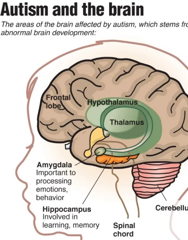
Autism is associated with widespread, developmentally dynamic changes in brain structure, connectivity, and cellular organization rather than a single “lesion.” These changes differ by age, brain region, and individual, but several patterns are now well supported.
## Overall brain growth trajectory
- Many children later diagnosed with ASD show **increased head circumference and total brain volume** in early childhood (roughly ages 1–4), especially in the first few years of life.
- This is often described as **early brain overgrowth**: excessive volume growth in infancy-early childhood, followed by a **slowing or plateau** in later childhood and, in some cases, a relative decline in adolescence-adulthood.
- However, only a **subset** of individuals show marked overgrowth; brain differences in ASD are heterogeneous.
## Gray matter and cortical structure
MRI studies consistently find region-specific differences in gray matter volume, cortical thickness, and surface area:
- **Cortical thickness**:
- In childhood, many studies report **increased cortical thickness** across frontal, temporal, parietal, and occipital regions.
- During adolescence and adulthood, there is often **accelerated cortical thinning**, particularly in frontal, parietal, and occipital lobes.
- **Surface area vs thickness**: In children, much of the increased brain volume in ASD appears driven more by **increased cortical surface area** than by thickness.
- **Regional gray matter**: Meta-analyses and voxel-based studies show both increases and decreases depending on region and age, including:
- **Decreases** in areas such as putamen, parts of the cerebellar vermis, hippocampus-amygdala complex, operculum, and some frontal and temporal regions.
- **Increases** in some frontal regions (medial prefrontal cortex, superior-inferior frontal gyri), middle temporal gyrus, and in certain perceptual areas.
These patterns align with core ASD features: social cognition, communication, and repetitive behaviors map onto altered structure in frontal, temporal, parietal, and subcortical regions.
## White matter and connectivity
White matter differences are a major theme in ASD neurobiology:
- **Volume and microstructure**:
- Early studies report **increased subcortical white matter**, especially in frontal lobes, and overall enlarged brain volume.
- Postmortem work shows **altered axonal density** and evidence of **impaired myelin** in white matter tracts.
- **Connectivity**: Diffusion tensor imaging (DTI) and functional MRI suggest **abnormal long-range connectivity** between brain regions, supporting models of disrupted integration across networks involved in social, language, and sensory processing.
## Subcortical structures: amygdala, hippocampus, basal ganglia, cerebellum
Several subcortical systems show consistent alterations:
- **Amygdala and hippocampus**:
- Postmortem studies report **smaller cell size and increased cell density** in the amygdala, hippocampus, limbic system, and entorhinal cortex across ages in ASD.
- Imaging studies often find **volumetric reductions** in clusters including the uncus-amygdala, parahippocampal gyrus, entorhinal cortex, and putamen, particularly in the right hemisphere.
- Some imaging work in children reports **increased amygdala-hippocampal volumes** early in development, highlighting age-dependent effects.
- **Basal ganglia (putamen, caudate)**:
- Meta-analyses identify structural anomalies in the basal ganglia and nearby regions, linked to repetitive behaviors and motor patterns.
- **Cerebellum**:
- One of the most consistent postmortem findings is a **reduction in Purkinje cell number and size**, especially in posterolateral neocerebellar and archicerebellar cortices.
- Imaging often shows **decreased cerebellar volume**, particularly in adolescents and adults.
## Cellular and microstructural changes
Beyond macroscopic imaging, neuropathology points to fine-grained developmental alterations:
- **Cortical cytoarchitecture**: Abnormalities in neuronal layering and organization in the cerebral cortex.
- **Cell size and density**:
- Smaller neurons with higher density in limbic regions and amygdala-hippocampus.
- Enlarged neurons reported in cerebellar nuclei and inferior olive in young patients.
- **White matter pathology**: Altered axonal packing and myelin integrity, consistent with connectivity deficits.
## Developmental perspective
ASD is best understood as a **disorder of neurodevelopmental trajectories.
- The “normal” trajectory of brain growth, neuronal patterning, and cortical connectivity is altered from early life.
- Structural differences evolve: early overgrowth and cortical thickening can transition to accelerated thinning and relative volume reductions in specific regions by adolescence-adulthood.
## Overall brain growth trajectory
- Many children later diagnosed with ASD show **increased head circumference and total brain volume** in early childhood (roughly ages 1–4), especially in the first few years of life.
- This is often described as **early brain overgrowth**: excessive volume growth in infancy-early childhood, followed by a **slowing or plateau** in later childhood and, in some cases, a relative decline in adolescence-adulthood.
- However, only a **subset** of individuals show marked overgrowth; brain differences in ASD are heterogeneous.
## Gray matter and cortical structure
MRI studies consistently find region-specific differences in gray matter volume, cortical thickness, and surface area:
- **Cortical thickness**:
- In childhood, many studies report **increased cortical thickness** across frontal, temporal, parietal, and occipital regions.
- During adolescence and adulthood, there is often **accelerated cortical thinning**, particularly in frontal, parietal, and occipital lobes.
- **Surface area vs thickness**: In children, much of the increased brain volume in ASD appears driven more by **increased cortical surface area** than by thickness.
- **Regional gray matter**: Meta-analyses and voxel-based studies show both increases and decreases depending on region and age, including:
- **Decreases** in areas such as putamen, parts of the cerebellar vermis, hippocampus-amygdala complex, operculum, and some frontal and temporal regions.
- **Increases** in some frontal regions (medial prefrontal cortex, superior-inferior frontal gyri), middle temporal gyrus, and in certain perceptual areas.
These patterns align with core ASD features: social cognition, communication, and repetitive behaviors map onto altered structure in frontal, temporal, parietal, and subcortical regions.
## White matter and connectivity
White matter differences are a major theme in ASD neurobiology:
- **Volume and microstructure**:
- Early studies report **increased subcortical white matter**, especially in frontal lobes, and overall enlarged brain volume.
- Postmortem work shows **altered axonal density** and evidence of **impaired myelin** in white matter tracts.
- **Connectivity**: Diffusion tensor imaging (DTI) and functional MRI suggest **abnormal long-range connectivity** between brain regions, supporting models of disrupted integration across networks involved in social, language, and sensory processing.
## Subcortical structures: amygdala, hippocampus, basal ganglia, cerebellum
Several subcortical systems show consistent alterations:
- **Amygdala and hippocampus**:
- Postmortem studies report **smaller cell size and increased cell density** in the amygdala, hippocampus, limbic system, and entorhinal cortex across ages in ASD.
- Imaging studies often find **volumetric reductions** in clusters including the uncus-amygdala, parahippocampal gyrus, entorhinal cortex, and putamen, particularly in the right hemisphere.
- Some imaging work in children reports **increased amygdala-hippocampal volumes** early in development, highlighting age-dependent effects.
- **Basal ganglia (putamen, caudate)**:
- Meta-analyses identify structural anomalies in the basal ganglia and nearby regions, linked to repetitive behaviors and motor patterns.
- **Cerebellum**:
- One of the most consistent postmortem findings is a **reduction in Purkinje cell number and size**, especially in posterolateral neocerebellar and archicerebellar cortices.
- Imaging often shows **decreased cerebellar volume**, particularly in adolescents and adults.
## Cellular and microstructural changes
Beyond macroscopic imaging, neuropathology points to fine-grained developmental alterations:
- **Cortical cytoarchitecture**: Abnormalities in neuronal layering and organization in the cerebral cortex.
- **Cell size and density**:
- Smaller neurons with higher density in limbic regions and amygdala-hippocampus.
- Enlarged neurons reported in cerebellar nuclei and inferior olive in young patients.
- **White matter pathology**: Altered axonal packing and myelin integrity, consistent with connectivity deficits.
## Developmental perspective
ASD is best understood as a **disorder of neurodevelopmental trajectories.
- The “normal” trajectory of brain growth, neuronal patterning, and cortical connectivity is altered from early life.
- Structural differences evolve: early overgrowth and cortical thickening can transition to accelerated thinning and relative volume reductions in specific regions by adolescence-adulthood.
Keywords